E. coli
Mastitis caused by Escherichia coli is usually an acute, sometimes even peracute (extremely fast progressing) disease with overall health impairment. The mammary gland is enlarged, solid, warm and very painful. Supramammary lymph nodes are significantly enlarged and painful at palpation. The cow suffers from fever, accelerated pulse, accelerated breathing and anorexia, it stops ruminating, can develop a toxaemia syndrome which results in lying down, shock and death.
Escherichia coli is a bacterium which multiplies very actively (even more often than every 20 minutes) in an appropriate environment (such as in the mammary gland), resulting in a very fast progress of the pathological process in the mammary gland. As early as 4 hours after infection the inflammatory reaction is fully developed and in the next 2 hours fever and general signs of overall health impairment occur.
Escherichia coli can be found in large numbers in the digestive system of cattle, other animals and humans. Until now, no strains were found to have a higher affinity for the mammary gland so basically any strain of the bacterium can be expected to be able to penetrate the udder and cause E. coli mastitis. Development of mastitis can be preceded by diarrhoea, e. g. connected with sudden change in feed, worse hygiene conditions, metabolism impairment etc. Especially high-yielding dairy cows and those with the decreased function of the teat canal closure are at risk of E. coli mastitis. E. coli can adhere very well to tissues thanks to short projections coming out of the bacterial cell surface (so called fimbriae) and then form a biofilm and spread into the udder in an ascending way. Interestingly, the urinary bladder infections in people often happen this way.
In most cases, the E. coli udder infection develops by three weeks after calving when teat canal sphincters do not function properly and the process of closure lasts up to 30 minutes compared to 10 minutes in the later phases of lactation.
Escherichia coli has a number of subtypes which differ on the level of aggression (toxin production, penetration into the udder). If the bacterium is washed out during the next milking, it does not form the biofilm and does not stay in the mammary gland but only somatic cell counts increase shortly (for several days) as a reaction to the toxin production and deviation in the milk conductivity.
Another option is the biofilm formation and multiplication in the udder, toxin production, epithelium (cellular lining) damage, development of mastitis = inflammatory reaction when even after the successful treatment the reparation of this state lasts for weeks.
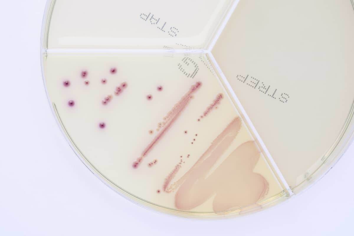
At the ClearMilk test, Escherichia coli grows in large pink colonies. It grows very well by 24 hours; it is essential to determine antibiotic susceptibility as quickly as possible to check the treatment already initiated because E. coli ranks among bacteria which often develop antibiotic resistance.
In the photo (above) from examination of milk from a cow with mastitis we can see that bacteria Escherichia coli and for example Streptococcus uberis can be differentiated unambiguously by colour and growth in one of the sectors of the dish; thus, we can have the first information about the causal agent of mastitis on a farm in 24 hours.